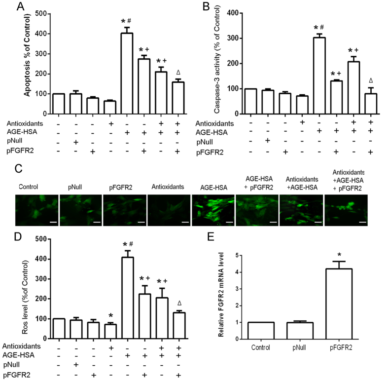
Figure 7. Effects of restoring FGFR2 on the protective effects of antioxidants against AGE-HSA-induced apoptosis in ADSCs.
The levels of apoptosis (A) and caspase-3 activity (B) were measured by ELISA. (C) Intracellular ROS generation was visualized under the fluorescence microscope. The scale bars represent 100 μm. (D) The level of DCF-sensitive ROS was measured by a flow cytometer.(E) Fored expression of FGFR2 was verified by RT-PCR.PReceiver-E2F3 or an empty pReceiver vector-transfected ADSCs were pretreated with antioxidants followed by treatment with AGE-HSA (300 μg/ml) for 24 h. Each value is expressed as the mean ± SD of three independent experiments. *P < 0.05 vs. control (HSA 300 μg/ml), #P < 0.05 vs. antioxidants (3 mM NAC and 0.2 mM AAP), +P < 0.05 vs. AGE-HSA (300 μg/ml), ΔP < 0.05 vs. antioxidants (3 mM NAC and 0.2 mM AAP) and AGE-HSA (300 μg/ml).

