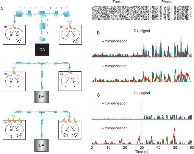
Figure 3.

Postsynaptic effects of DA cell firing. A: Schematic illustration of hypothetical postsynaptic adaptations to passive stabilization. A, top: DA signals in D1 and D2‐receptor expressing GABAergic medium spiny striatal projection neurons (SPNs) under normal conditions. Cyan indicates the DA level produced by alternation of phasic (P) and tonic (T) DA neuron firing. Dots inside D1‐ and D2‐SPNs represent the products of down‐stream signaling, for example, cAMP levels. These products are mainly produced under phasic DA signals, and we assume that they provide negative feedback on the receptor‐effector unit (orange). A, middle: Partial denervation has reduced the amplitude of phasic DA levels (cyan) and the activation of postsynaptic cascades is reduced. A, bottom: Increasing the gain of postsynaptic receptors phasic signals can compensate reduced phasic signals, but modeling shows increased activation under tonic firing. B and C: Implementation of adaptive pathways in the computational model. Blue, intact signaling; green, 75% denervated; red, 97% denervated. The raster plot is the same as in Figure 2A. B, upper: D1 receptor mediated signaling without compensation. In the absence of compensation, the postsynaptic activation during phasic firing is decreased by denervation. B, bottom: Denervated D1 signal with phasic amplitude compensation. C: Same as B, but for D2 signaling. [Color figure can be viewed in the online issue, which is available at wileyonlinelibrary.com.]
