
Figure 4.
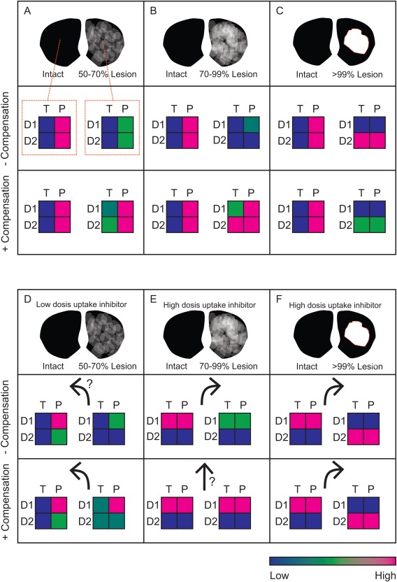
Overview of predictions for unilateral lesions from the theoretical analysis in Dreyer 6. Striatal sections with unilateral lesions are divided into 3 cases: low coherent (A, D), high coherent (B, E), and void (C, F). Activation level of D1‐ or D2‐regulated signals (D1 and D2, respectively) under tonic (T) and phasic (P) signaling are indicated by the color scale. A, B, and C are under normal conditions, whereas D is under low dopamine uptake inhibition. E and F are under high‐dose psychostimulant challenge. Arrows indicate predicted animal rotation direction (relative to the lesion). [Color figure can be viewed in the online issue, which is available at wileyonlinelibrary.com.]