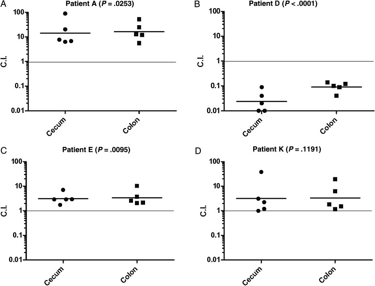
Figure 3.
Related persistent isolates differ in their pathogenicity in vivo. Groups of 5 female C3H/HeNHsd mice were treated with streptomycin and 24 hours later were infected orally with a 1:1 mixed inoculum of early and later S. Typhimurium isolates (5 × 10⁶–1 × 10⁷ CFU from each isolate) obtained from patients A (isolate 85982 and 87541; panel A), D (102261 and 102923; panel B), E (128781 and 130302; panel C), and K (135497 and 137309; panel D). Four days post infection mice were sacrificed, and the colonization of the later isolates relative to the early isolate was determined in the cecum and colon as explained in “Materials and Methods” section. Each point shows the competitive index (C.I.) value in a single mouse, whereas the geometrical mean of each group is indicated by the horizontal line. Two-tailed 1-sample t-test against a theoretical mean of 1.00 was used to determine statistical significance and is shown in brackets. Abbreviation: CFU, colony-forming unit.

