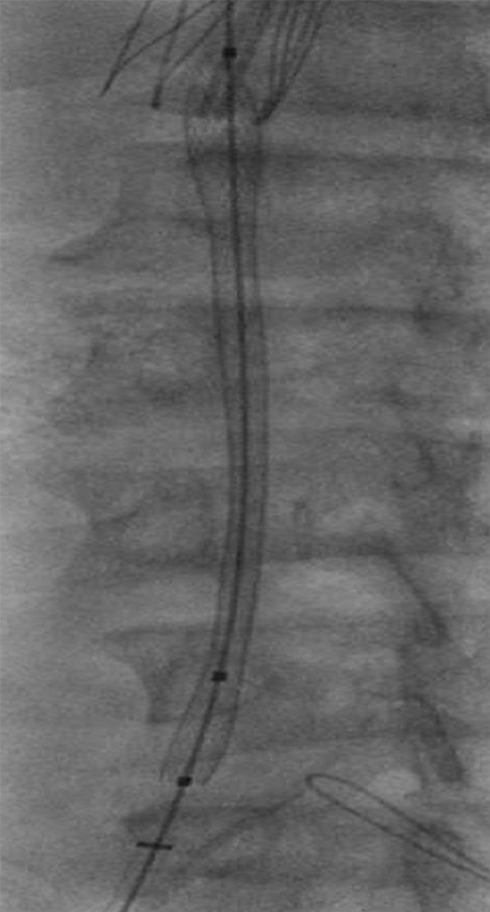
Figure 11e:

Creation of a neo-IVC 17 years after traumatic laceration. (a) Transjugular IVC contrast material injection demonstrates no detectable infrarenal IVC. (b) Right iliac venogram shows numerous collaterals and diminutive native vein. (c) Spot fluoroscopic image demonstrates a snare within a sheath that has been advanced through sharp recanalization (arrow) and two wires via bilateral common femoral veins (arrowheads). The right iliac wire has entered a lumbar collateral (based on subsequent imaging) and is not in the native IVC. (d) Spot fluoroscopic image of the snare cinching the back end of a stiff wire within the retroperitoneal fat. (e) Fluoroscopic image of undilated self-expanding stainless steel stent spanning the length of the absent IVC. (f, g) Digital subtraction venograms demonstrate brisk flow through the iliac and IVC stents into the suprarenal IVC and right atrium. See also Figs E2a–E2d in this patient (online). (Case courtesy of Thomas Sos, MD, and Akhilesh Sista, MD, Weill Cornell Medical College.)
