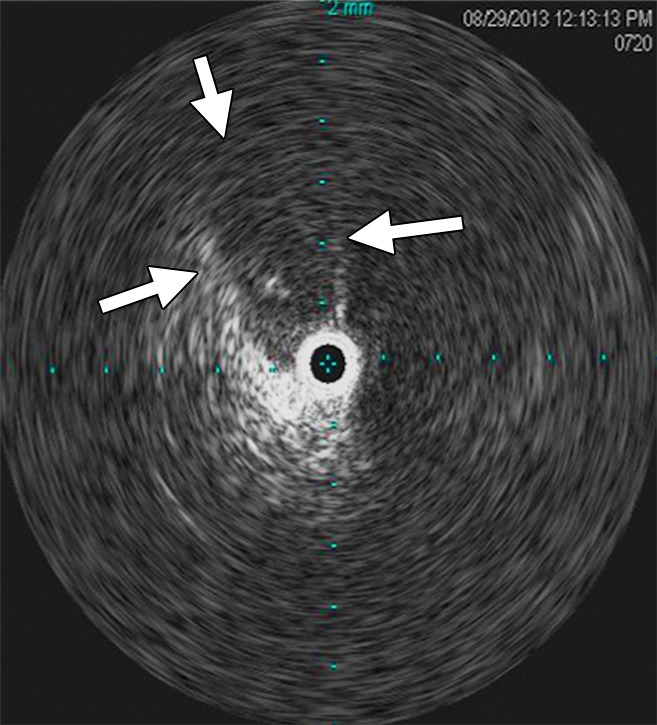
Figure 13b:

Intravascular US before and after stent placement. (a) Left iliac venogram demonstrates an occlusive lesion in the peripheral external iliac vein (arrow). (b) Intravascular US image at the level of compression. The arrows delineate the estimated borders of the narrowed venous segment. (c) Intravascular US image of the common iliac vein just central to the stenosis in image b. (d) Venogram obtained after stent placement demonstrates markedly improved flow and caliber of the left pelvic deep veins. (e) Intravascular US image obtained after stent placement at the level of the stenosis in image b reveals the improved caliber and the hyperechoic stent in cross section.
