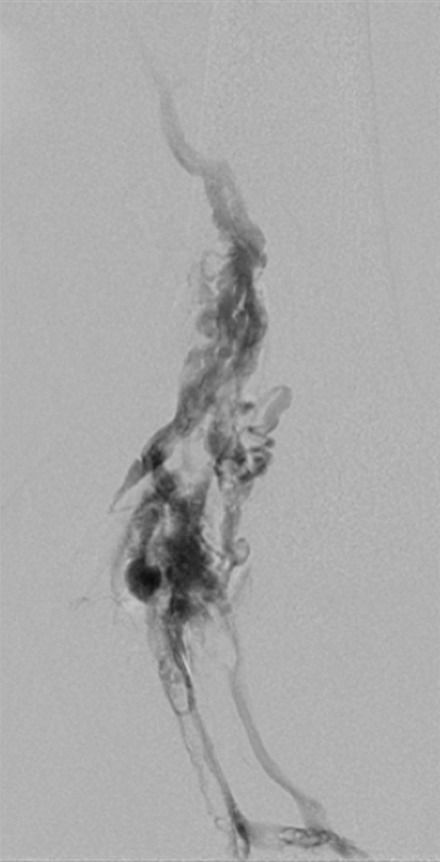
Figure 15b:

Recanalization of chronically occluded left femoropopliteal veins. (a) Spot fluoroscopic image of wire access into the left posterior tibial vein at the level of the ankle. (b) Popliteal venogram demonstrates heavily diseased popliteal vein, expanded by chronic thrombus, with partially recanalized channels. (c) Femoral venogram shows atretic femoral vein with collateralization. (d) Prolonged balloon angioplasty of the central femoral vein, with a representative waist at areas of stenosis. (e–g) Venograms obtained after angioplasty show improved flow and resolution of collaterals through the (e) popliteal, (f) peripheral femoral, and (g) central femoral veins. Three months later, the patient had no objective PTS per Villalta assessment, and (h) duplex US demonstrated a patent femoral vein. See also Figs E4a and E4b in this patient (online).
