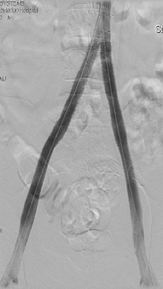
Figure 1e:

CDT and stent placement in a patient with progressive bilateral DVTs in spite of anticoagulation. (a) Left femoral venogram (patient prone) demonstrates extensive acute thrombus along the length of the vein. (b) Right iliac venogram demonstrates no filling of the iliac vein. (c) Fluoroscopic image depicts infusion catheters along the length of the left and right iliac thrombi. (d) Postinfusion left femoral venogram demonstrates excellent patency. (e) After stent placement, venogram of both iliacs demonstrates rapid flow through the stents (see also Figs E1a and E1b in this patient [online]).
