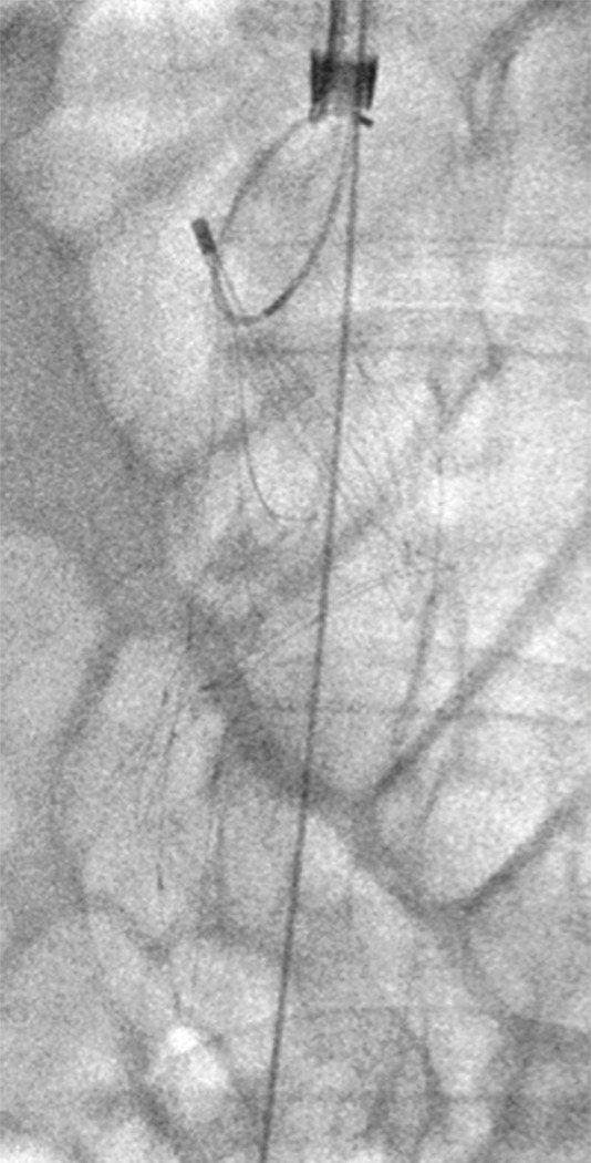
Figure 3d:

Caval thrombus treated with large-bore aspiration device. (a) IVC venogram demonstrates extensive caval thrombus and a malpositioned suprarenal IVC filter. (b) Fluoroscopic image depicts the suction/aspiration device (AngioVac; Angiodynamics) in the IVC. The arrow points to the balloon at the tip, which when inflated flares the tip. (c) Photograph of the recirculation filter shows bulky extracted thrombus. (d) Fluoroscopic image during filter retrieval shows a tip deflecting wire grasping the malpositioned filter. The tip of the wire has been snared, and the filter is subsequently pulled through the sheath. (e) IVC venogram obtained the next day after IVC filter removal demonstrates marked reduction in thrombus burden and free flow through the cava. No lytic drug was used during this case owing to a hemorrhagic stroke in this patient 3 weeks earlier.
