
Figure 8b:
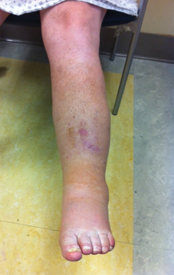
MR appearance of May-Thurner variant. (a) On the high-spatial-resolution blood-pool steady-state axial MR image, the left common iliac vein is compressed (arrowhead) between the left internal iliac artery and vertebral body. (b) The patient’s left leg has altered pigmentation, swelling, and an ulcer overlying the anterior tibia.