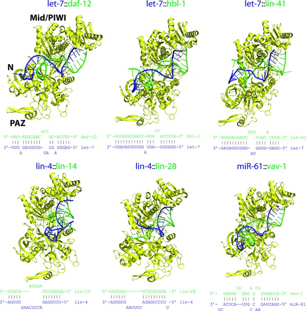
Figure 6.

Models of C. elegans miRISC structures. All viable structures were assembled using the method outlined in Figure 1; each displayed structure is ranked in the top 5. Complexes were derived from ALG-2 and a set of miRNA-target duplexes (secondary structures shown) from experimental literature; the initial ALG-2 structure (without the unstructured residues 1–73) was modeled using human AGO1 and AGO2 templates via comparative modeling. Color scheme: Ago, yellow; guide RNA strand, blue; and mRNA strand, green.
