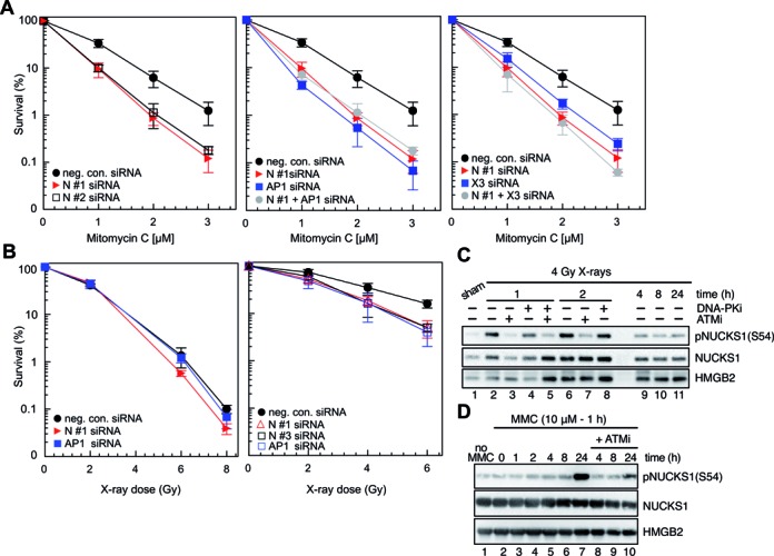
Figure 2.

NUCKS1 knockdown sensitizes HeLa cells to mitomycin C (MMC) and X-rays, and NUCKS1 is phosphorylated after DNA damage. (A) Survival curves obtained in control cells (transfected by neg. con. siRNA) or after NUCKS1 depletion following transfection with either NUCKS1 siRNA N #1 or NUCKS1 siRNA N #2 after treatment with MMC (left panel). NUCKS1- and/or RAD51AP1-depleted HeLa cells show the same increased cellular sensitivity to MMC treatment compared to control transfected cells (middle panel). NUCKS1- and/or XRCC3-depleted HeLa cells show similarly increased cellular sensitivity to MMC treatment compared to control transfected cells (right panel). Data points are the means from 3 independent experiments ± 1SD. (B) Cell survival curves obtained after exposure of asynchronous HeLa cells with NUCKS1 or RAD51AP1 depletion to graded doses of X-rays (left panel). Cell survival curves obtained after exposure of HeLa cells in S-phase and with NUCKS1 or RAD51AP1 depletion to graded doses of X-rays (right panel). Data are the mean from 3 independent experiments ± SD. (C) Western blots of PCA extracts obtained from HeLa cells to show that NUCKS1 is phosphorylated in an ATM-dependent manner on serine 54 (S54) after exposure to X-rays. HMGB2 serves as loading control. (D) Western blots of PCA extracts obtained from HeLa cells to show that NUCKS1 is phosphorylated by ATM on serine 54 (S54) after exposure to MMC. HMGB2 serves as loading control.
