
Figure 1.
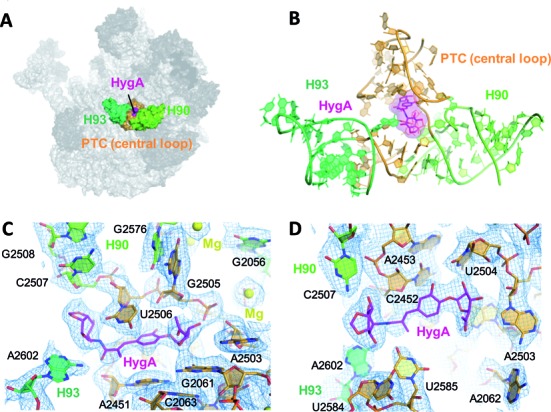
The HygA binding site. (A) An overview showing the location of HygA (colored in magenta) on the Deinococcus radiodurans 50S ribosomal subunit. (B) The HygA binding site is formed by 23S rRNA helices H90 and H93 (light and dark green, respectively) and the PTC central loop (orange). (C and D) Final 2Fo-Fc electron density map sharpened, contoured at 1 sigma and shown from two different perspectives.