
Figure 3.
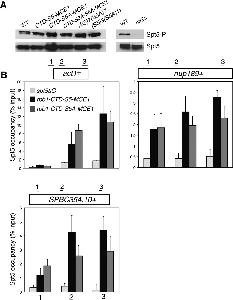
Loss of Rpb1 Ser5 does not impair Spt5 phosphorylation or its association with transcribed chromatin. (A) Whole-cell extracts derived from the indicated strains (top) were subjected to SDS-PAGE and immunoblotting with the indicated antibodies (right). ‘WT’ in the left panel is as in Figure 1; ‘WT’ in the right panel is as in Figure 2. (B) ChIP of Spt5 was carried out using a polyclonal antibody recognizing the Spt5 CTD in the indicated strains. A spt5ΔC strain lacking the entire CTD served as a negative control. ChIP signals were quantified by qPCR using primer pairs at the numbered locations across the act1+, nup189+ and SPBC354.10+ genes as shown and expressed as% of signal in the whole-cell extract (input). Error bars denote standard deviations from three independent experiments.