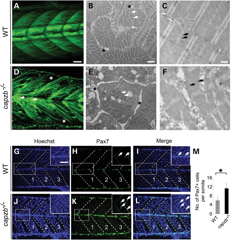
Figure 4.
capzb mutants show muscle disorganization. Confocal images of somites using Tg(acta1a:lifeact-GFP) expressing GFP in skeletal muscles under the control of an α-actin promoter reveal disorganization of the muscles in capzb mutants (D, asterisk, arrows), but not in wild-type embryos (A). Ultrastructure examination by TEM reveals extensive loss of muscle fibers (E, arrowheads) within the fascicles (bundle of muscle fibers) with a large perimysial space (E, asterisk) in mutants compared with the wild-type (B), in transverse cross-sections. Examination of longitudinal sections of trunk skeletal muscles reveals loss of structural integrity and disorganization of the sarcomeres in capzb mutants (C, arrowheads) compared with wild-type embryos (F). Pax7 immunohistochemistry on 65 hpf wild-type (n = 3) and capzb mutants (n = 3) (G–L). Somite number 13–15 (marked 1, 2, 3) was imaged in all samples, in blue (Hoechst) and green (Pax7) channels. Insets in each image show the region-of-interest (white boxes) in higher magnification. Arrowheads in the green and merge images depict both Pax7+ and Hoechst+ cells, which were counted for quantification. (M). The average number of Pax7+ and Hoechst+ cells/somite in 65 hpf capzb mutants (n = 9) is increased compared with the wild-type (n = 9). Somite number 13–15 (marked 1, 2, 3) was counted. *P < 0.0001. Scale bars: 20 μm (A, D, G–L), 2 μm (B and E), 500 nm (C and F), 5 μm (insets, G–L).

