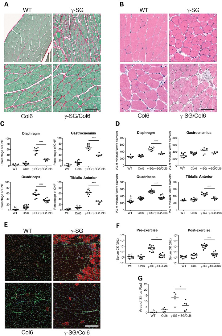
Figure 3.
Muscle pathology is decreased in γ-SG-null/Col6a2Δex5 mice. (A) Representative micrographs of Sirius red-stained quadriceps muscles from WT, Col6, γ-SG and γ-SG/Col6 mice (14–20 weeks). (B) Representative micrographs of H&E-stained quadriceps muscles from WT, Col6, γ-SG and γ-SG/Col6 mice (14–18 weeks). (C) Quantification of the percentage of regenerating muscle fibers (CNFs, centrally nucleated fibers) in muscles from WT, Col6, γ-SG and γ-SG/Col6 mice (14–18 weeks). ****P < 0.0001. (D) Quantification of fiber size variation (VC = variance coefficient; 1000 × standard deviation of minimal Feret's diameter/mean of minimal Feret's diameter) in muscles from WT, Col6, γ-SG and γ-SG/Col6 mice (14–18 weeks). ****P < 0.0001. (E) Representative micrographs of EBD uptake (red) and perlecan (green) immunofluorescence staining in quadriceps muscles from WT, Col6, γ-SG and γ-SG/Col6 mice (12–16 weeks) after exercise. (F) Serum CK levels in WT, Col6, γ-SG and γ-SG/Col6 mice (10–12 weeks), both at baseline (pre-exercise) and 2 h after downhill exercise (post-exercise). **P < 0.01 and ****P < 0.0001. (G) Quantification of Sirius red-stained quadriceps muscles from WT, Col6, γ-SG and γ-SG/Col6 mice (14–20 weeks). *P < 0.05. Samples shown in (A, B and E) were prepared and processed simultaneously, and images were taken at the same exposure level. Scale bar: 100 μm (A and B) or 200 μm (E). Closed circles represent male mice, and open circles represent female mice in (C, D, F and G).

