Figure 3.
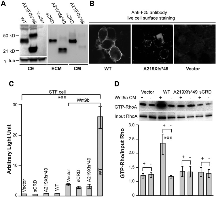
FZD5-A219Xfs*49 is incapable of mediating Wnt signaling. (A) Immunoblot analysis of subcellular fractions from transfected HEK293 cells. FZD5 A219Xfs*49mutant protein is detected primarily in ECM fraction. sCRD is expressed in both the culture medium and ECM. CE, cell extract. (B) Live cell immunofluorescence detection. Immunofluorescence staining was conducted to detect FZD5 proteins expression in transfected cells on coated coverslips (see the ‘Materials and Methods’ section). WT FZD5 is primarily present on the cell surface (left panel), while the majority of A219Xfs*49 mutant protein is detected extracellularly (middle panel), presumptively in ECM (doted staining). Negative control with vector transfection is shown in the right panel. (C) Wnt9b-induced canonical Wnt signaling in STF reporter cell line. Cells were transfected with 0.5 µg Wnt9b plasmid combined with 0.25 µg other plasmids. Like sCRD, FZD5 A219Xfs*49 mutant protein is not able to mediate Wnt9b-induced canonical Wnt signaling. The rightmost bar represents Wnt9b-induced canonical Wnt activity by WT FZD5, which is significantly different from all other forms of FZD5. (D) Representative image for active-Rho pulldown assays for non-canonical Wnt signaling. HEK293 cells were transfected with FZD5 WT, A219Xfs*49 and sCRD plasmids, treated with Wnt5a recombinant protein conditioned medium. Active GTP-RhoA assays strictly followed the manufacturer instructions (Cytoskeleton, Inc., Cat. no. Bk-030) (for details see the ‘Materials and Methods’ section). Wnt5a-enhanced formation of GTP-RhoA is obtained in the presence of FZD5, but not A219Xfs*49 mutant or sCRD. The signal intensities were quantified by NIH ImageJ from three immunoblots for three independent experiments, and analyzed by the Microsoft Excel (Student's t-test, ***P < 0.001).

