Figure 5.

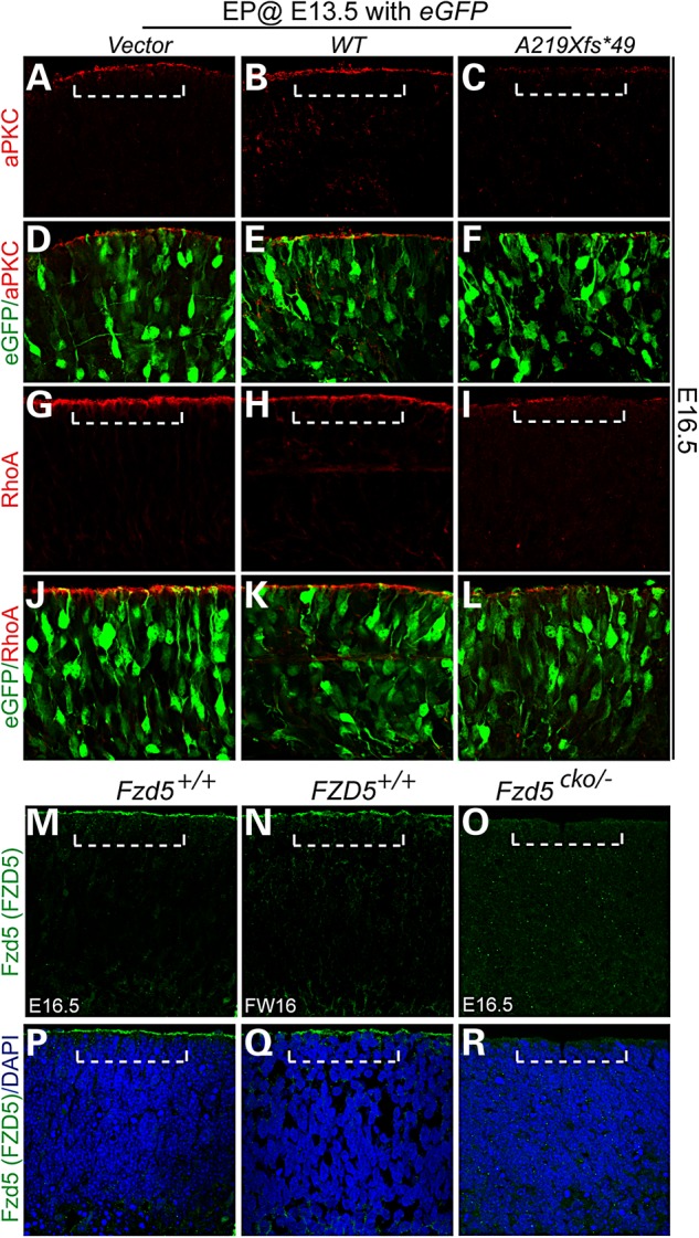
Overexpression of FZD5 A219Xfs*49 led to similar apical junction defects that were reported in mouse Fz5/8 compound mutants. Mouse embryonic (E13.5) retina was dissected and subjected to electroporation supplied with WT FZD5 and FZD5 A219Xfs*49 DNA solution. The retinae were cultured for 72 h and harvested for immunohistochemistry. (A–F), aPKC localization in vector (A), WT FZD5 (WT) (B) and A219Xfs*49 mutant (C) electroporated retinae. Note the loss of apical localization of aPKC in A219Xfs*49-expressing retina (C). (D–F) Images of (A–C) merged with co-elctroporated eGFP, respectively. (G–I) Similar as aPKC, apical RhoA enrichment is also greatly attenuated (compared with G and H). (J–L) Images of (G–I) merged with co-elctroporated eGFP, respectively. (M–R) Localization of FZD5 protein in mouse and human retina. (M) Apical localization of the FZD5 protein in WT mouse retina (above dashed bracket). (N) Same protein localization of FZD5 was detected in human retina. (O) Mouse Fzd5 conditional mutant retina showed the absence of apical FZD5 protein. (P–R) Images from (M–O) merged with 4′,6-diamidino-2-phenylindole, respectively.
