Figure 6.
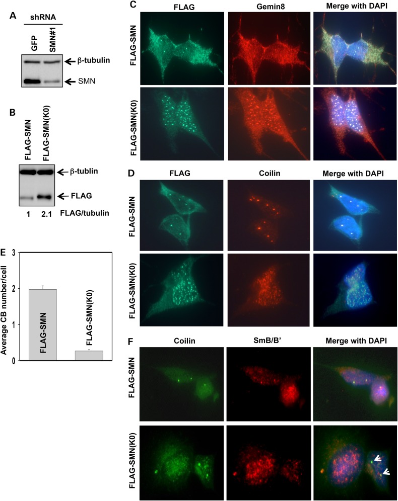
SMN(K0) primarily resides in the nucleus, and expressing SMN(K0) impairs canonical CBs and coilin/snRNP co-localization. (A) Knockdown of SMN in SH-SY5Y cells using the shRNA construct that targets the 3′ UTR of the SMN1 gene. An shRNA targeting GFP was used as a negative control. Whole-cell lysates were immunoblotted for SMN and β-tubulin. (B) Stably overexpressing FLAG-SMN or FLAG-SMN(K0) in SMN knockdown SH-SY5Y cells. Whole-cell lysates were used for immunoblotting of FLAG and β-tubulin. Protein levels were quantitated by desitometry, with the level of FLAG-SMN being referenced as 1. (C) Co-immunostaining of FLAG and Gemin 8 in SMN knockdown SH-SY5Y cells stably expressing FLAG-SMN or FLAG-SMN(K0). (D) Co-immunostaining of FLAG and coilin in SMN knockdown SH-SY5Y cells stably expressing FLAG-SMN or FLAG-SMN(K0). (E) Quantification of canonical CBs (>0.3 µm in diameter) in SMN knockdown SH-SY5Y cells stably expressing FLAG-SMN or FLAG-SMN(K0). Three different areas of each staining were chosen for CB counting (50 cells/area). Error bars represent SD. (F) Co-immunostaining of SmB/B′ and coilin in SMN knockdown SH-SY5Y cells stably expressing FLAG-SMN or FLAG-SMN(K0). Arrows denote CBs that did not co-localize with Sm.

