
Fig. 10.
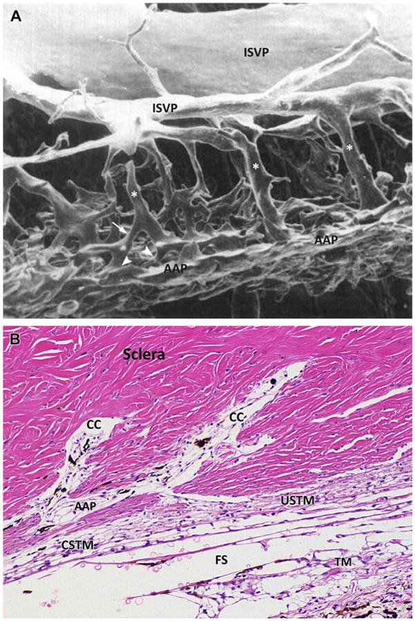
(A) Luminal cast of the canine trabecular outflow pathway. The vessels of the outflow at the level of the angular aqueous plexus (AAP) display a complex and tangled network made by smaller channels oriented tangentially in different planes and directions. Larger radial collector channels (asterisks) connect the network to larger venous vessels in the outer sclera, the intrascleral venous plexus (ISVP). Radial channels are displaced at different anterior and posterior sites. Irregular patterns of anastomoses are present among all the channels and shorter radial channels are seen (arrow). The connections between almost perpendicular inner and radial channels are called ostia (arrowheads and Fig. 7). (B) Microphotograph of a histologic sagittal section of the inner scleral region at the level of the scleral sulcus in a normal canine eye showing the complex anastomotic pattern of the drainage system in dogs. Hematoxylin and eosin staining. Two large, radial collector channels (CC) are visible anteriorly and posteriorly. These collector channels convey the aqueous filtered through the complex system represented by trabecular meshwork (TM), CSTM, uveoscleral TM (USTM), and angular aqueous plexus (AAP). Fontana spaces (FS) are present between the beams of the TM. (Adapted from Van Buskirk EM. The canine eye: the vessels of aqueous drainage. Invest Ophthalmol Vis Sci 1979;18:226; with permission.)