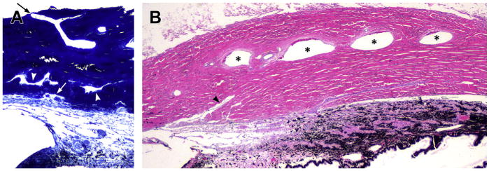
Fig. 11.

The scleral outflow systems. (A) The complex angular aqueous plexus (AAP) and collector channels are displaced in different layers with different directions and different sizes in the inner sclera. The arrowheads indicate collector channels. The white arrow points to an ostium between AAP and a short radial collector channel. In the outer sclera an aqueous vein can be seen directed to the limbus and to the anterior ciliary veins (black arrow) (toluidine blue). (B) Longitudinal section of the sclera corresponding to the ciliary body area. Four large scleral veins representing the intrascleral venous plexus (ISVP) are indicated with asterisks. The arrowhead points to a large, radial collector channel directing to the ISVP area (Hematoxylin and eosin).
