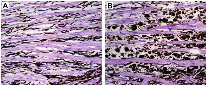
Fig. 12.

Photomicrographs of ciliary body sections posterior to the trabecular meshwork in 2 normal eyes from different dogs. (A) Ten month old male Labrador. The longitudinal bundles of the ciliary muscle alternate with connective tissue mostly filled with fusiform dendritic melanocytes representing the normal pigmented component of uveal ocular tissue. (B) Fifteen year old spayed female Labrador. The intermuscular spaces present a large number of plump large pigmented cells representing infiltrating macrophages melanin-laden (melanophages).
