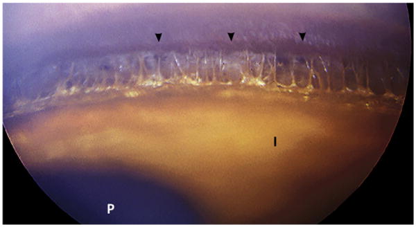
Fig. 3.

Normal iridocorneal angle in a dog at gonioscopy. Lightly pigmented iris tissue strands can be observed spanning the peripheral angular space and introducing to the posterior space of the ciliary cleft. The corneoscleral pigmented line (Schwalbe's line) is indicated by the arrowheads. I, anterior surface of the iris; P, pupil.
