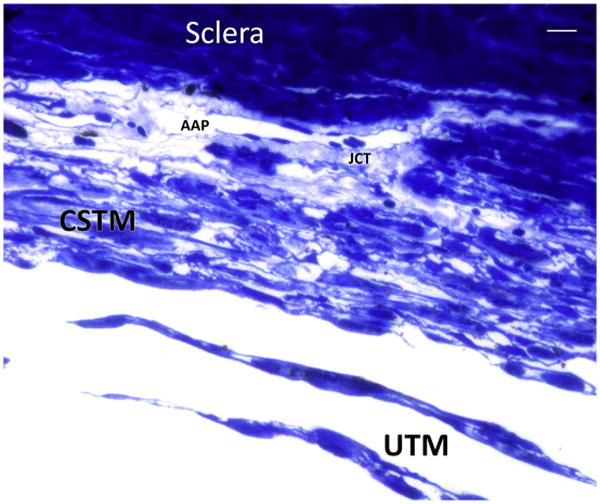
Fig. 8.

Semithin section with toluidine blue staining shows the transition between the uveal trabecular meshwork (UTM) represented by irregularly distributed beams of connective tissue lined by endothelial cells into a more lamellar and organized tissue represented by the corneoscleral trabecular meshwork (CSTM). Small, interlamellar spaces can be seen. The justacanalicular connective tissue (JCT) is a thin layer rich in extracellular matrix that surround the inner wall of the endothelial cells of angular aqueous plexus (AAP). Bar 5 10 μ.
