
Fig. 9.
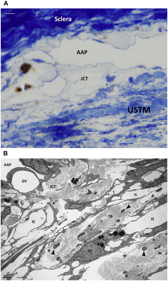
(A) Semithin section with toluidine blue staining of the angular aqueous plexus (AAP) region in a canine eye. The lamellar organization of the uveoscleral trabecular meshwork (USTM) can be recognized at the bottom. Some interlamellar spaces can be seen. The transition region between the USTM and the AAP is represented by a loose translucent tissue, the juxtacanalicular connective tissue (JCT). Bar 5 5 μ. (B) Transmission electron microscopy. A microphotograph of a same corresponding area shows the trabecular beams (TB) of the USTM. The beams are constituted by collagen fibers and larger elastin fibers (arrowheads). Endothelial trabecular meshwork cells line the beams and engulf melanin granules (EC). Intertrabecular spaces (IS) are reducing toward the JCT. A giant vacuole (GV) can be seen within the cytoplasm of an endothelial cell of the inner wall of the angular aqueous plexus (AAP).