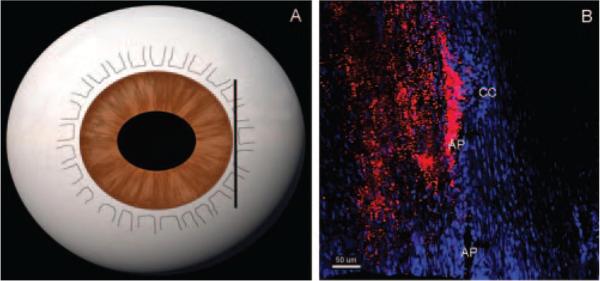
Figure 5.
Tracer distribution in frontal sections at 15 mm Hg. In frontal sections, tangential to the corneoscleral limbus and perpendicular to the ocular surface (A), segmental tracer distribution concentrated near the CC ostium was observed (B). Less tracer labeling was seen along the AP away from CC ostium. Scale bar, 50 μm.

