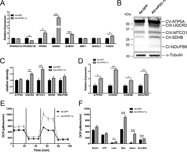
Figure 4.
Overexpression of mPGC-1α in hfRPE increases transcription factor and OXPHOS gene expression, and oxidative metabolism. (A) Gene expression analysis by qPCR of PPARGC1A and PPARGC1B and selected transcription factors 48 hours following ad-mPGC-1α infection of hfRPE. (B, C) Representative Western-blot (B) and quantification of OXPHOS proteins (C) shows increased expression of UQCR2 and MTCO1 in hfRPE 48 hours after adenovirus infection (n = 3 each group). (D) Gene expression analysis confirmed the induction of multiple OXPHOS genes in hfRPE (n = 3 each group). (E) Oxygen consumption rates in GFP-infected (black line) and mPGC-1α–infected (gray line) hfRPE. Oligomycin (1 mM), FCCP (500 nM), and rotenone and antimycin A (2 mM) were injected following the third, sixth, and ninth measurement, respectively. (F) Analysis of OCR parameters. PGC-1α significantly increased multiple phases of respiration in hfRPE (n = 5 each group).

