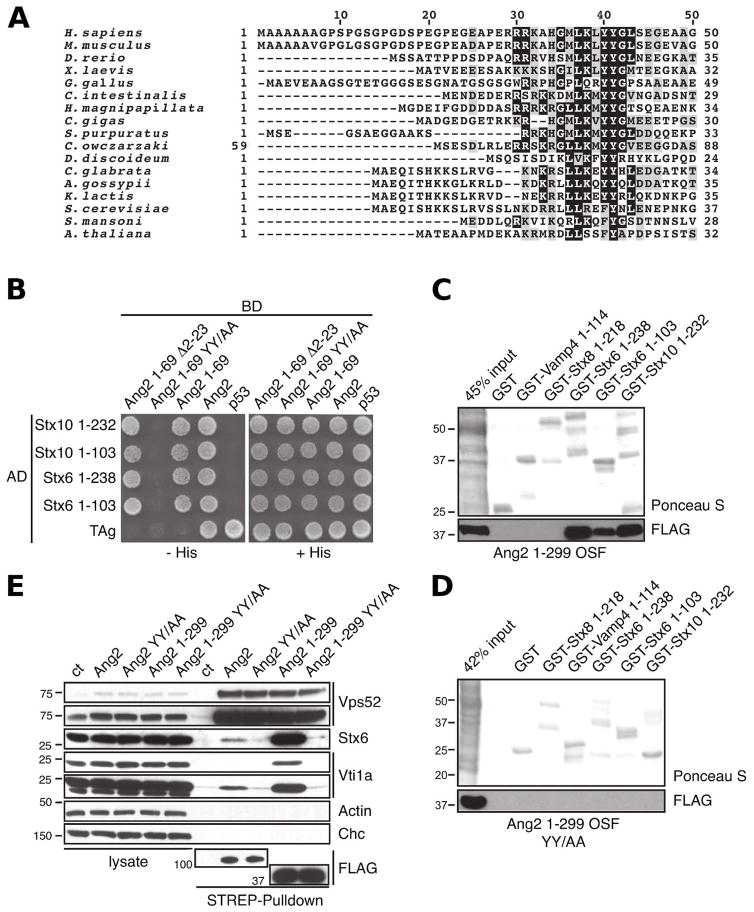
Figure 2.
A di-tyrosine motif in Ang2 is required for binding to the Habc domain of Stx6 and Stx10. (A) Alignment of N-terminal segments of Ang2/Vps51 from different species using ClustalW. (B) Y2H analysis of the interaction of Stx6 and Stx10 fragments with full-length Ang2, Ang2 1-69, Ang2 1-69 with a deletion of amino-acids 2-23 (Ang2 1-69 Δ2-23) and Ang2 with the conserved di-tyrosine motif mutated to alanine (Ang2 1-69 YY/AA). (C) In vitro binding of Ang2 1-299 OSF or (D) Ang2 1-299 OSF YY/AA expressed in HeLa cells to GST-SNAREs. The experiment was performed as described in the legend to Fig. 1D. (E) StrepTactin-pulldown assay of endogenous SNAREs with different Ang2 OSF constructs expressed by transfection in HeLa cells. Endogenous Stx6 and Vti1A SNAREs as well as clathrin heavy chain (Chc) and actin (loading controls) were detected by immunoblotting The positions of molecular mass markers (in kDa) are indicated on the left of panels C, D and E.

