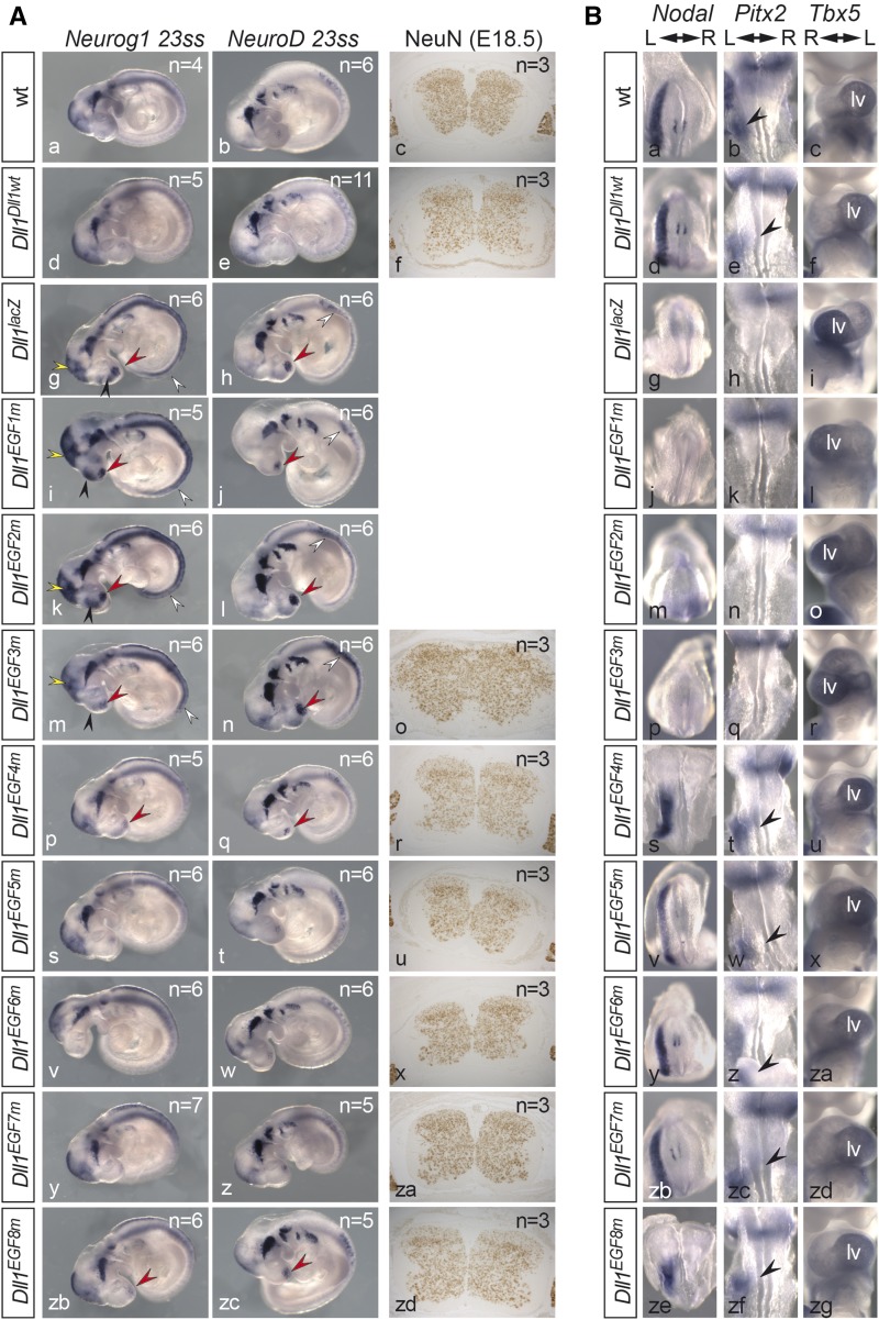
Figure 4.
Neuronal differentiation and left–right asymmetry in mutants homozygous for individual EGF alleles. (A) WISH of E9.5 and antibody staining of spinal cord sections of E18.5 embryos. Alleles are indicated at the left, analyzed markers at the top. Arrowheads point to regions of upregulated gene expression in the nasal placode (red arrowhead), midbrain (yellow arrowhead), forebrain (black arrowhead), and spinal cord (white arrowhead). For NeuN, we analyzed three individual embryos with a minimum of three and a maximum of nine consecutive sections per genotype. (B) WISH of six ss (Nodal, dorsal views) E8.5 (Pitx2, dorsal views) and E10.5 (Tbx5, ventral views) embryos. Alleles are indicated at the left, probes at the top. For genotyping of Dll1EGF4m (s) and Dll1EGF8m (ze) 6 ss embryos, the posterior halves of embryos were removed prior to hybridization to Nodal. Black arrowheads in (b, e, t, w, z, zc, zf) point to Pitx2 expression in the left LPM. “lv” indicates the Tbx5-positive ventricle normally positioned on the left side. The numbers of analyzed embryos are summarized in Table S6 and Table S7.

