
Fig 4. Grem1 expression during mouse craniofacial development.
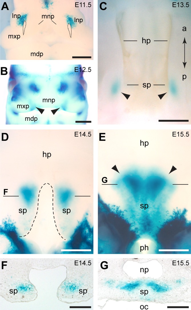
(A-E) Expression of Grem1 is visualized by X-Gal staining of heterozygous Grem1LacZ whole mount embryos. (A) At E11.5, Grem1 is expressed in the dorsal part of the lateral nasal prominence (lnp). Stippled lines demarcate the nasal pits. (B) At E12.5, Grem1-positive domains are also detectable in the merging zones (arrowheads) of medial nasal prominences (mnp) and maxillary prominences (mxp). (C-G) Secondary palate development. (C) At E13.5, Grem1-positive domains are observed in the forming soft palate (sp). (D) At E14.5, the hard palate (hp) has formed while the Grem1-expressing shelves of the soft palate are not yet fused. (E) At E15.5, the soft palate has fused and Grem1 expression extends posterior to the pharynx (ph). Note the sharp anterior boundary of Grem1 expression in the soft palate (arrowheads). (F, G) Sections of whole mount stained embryos. (F) Cross section at the level indicated in (D) showing that Grem1 expression is restricted to the mesenchyme. (G) Cross section at the level indicated in (E) showing Grem1 expression in the soft palate, which separates the nasopharynx (np) from the oral cavity (oc). Additional abbreviations: a, anterior; l, lateral; m, medial; mdp, mandibular prominence; p, posterior. Scale bars: 500μm.