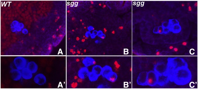
Figure 9.
Female germ cells compromised for sgg function undergo premature mitosis. Both wild type and embryos derived from females carrying sgg germline clones were probed with Sxl (not shown), Vasa (blue) and Phospho-histone H-3 (red) antibodies. Embryos in A–C show female embryos of the denoted genotype. Wild-type stage 14 embryo in A is devoid of pH 3 specific staining (magnified view of just the PGCs in A′) whereas sggm- germ cells shown in panels B (stage 14) and C (stage 14) display pH 3. B′ and C′ show magnified view of just the PGCs from the same embryos with a clear presence of pH 3 specific signal. For quantitation see Table 1. Embryos between stages 13 and 15 were used for analysis.

