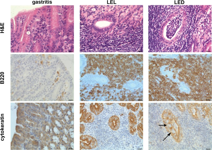
Fig 1. Histology and immunohistochemistry of gastric pathology.
Histological and immunohistochemical staining of representative cases of chronic gastritis with lymphoid aggregates and MALT lymphoma with lymphoepithelial lesions (LEL) or lymphoepithelial destruction (LED); H&E staining, B220 labeling B-cells and anti-cytokeratine staining labeling the epithelium. Centrocyte-like cells infiltrate the gastric epithelium (arrows). Scale bars show 20 μm. Original magnification x40.

