Figure 5.

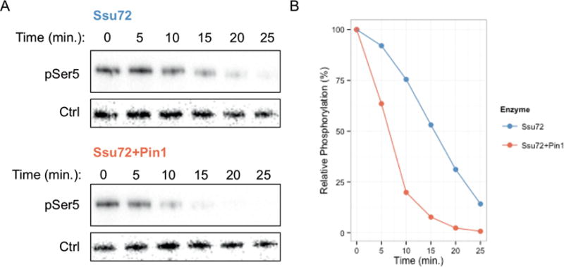
In vitro reconstruction Pin1 mediated Ssu72 enhancement. (A) Western blot against TFIIH phosphorylated GST-CTD dephosphorylated by Ssu72 with or without Pin1. Phospho-Ser5 (pSer5) was monitored for reactions containing Ssu72 alone (top) or Ssu72 + Pin1 (bottom) over the indicated time course. Mouse IgG heavy chain (Ctrl) was introduced during reaction quenching to provide a loading control. (B) Quantification of Western blot. Phospho-Ser5 bands were first normalized to loading control and then relative to the respective zero time point for each condition. Blot and quantification represent one experimental replicate of three independent experimental replicates, all displaying increased dephosphorylation upon Pin1 supplementation.
