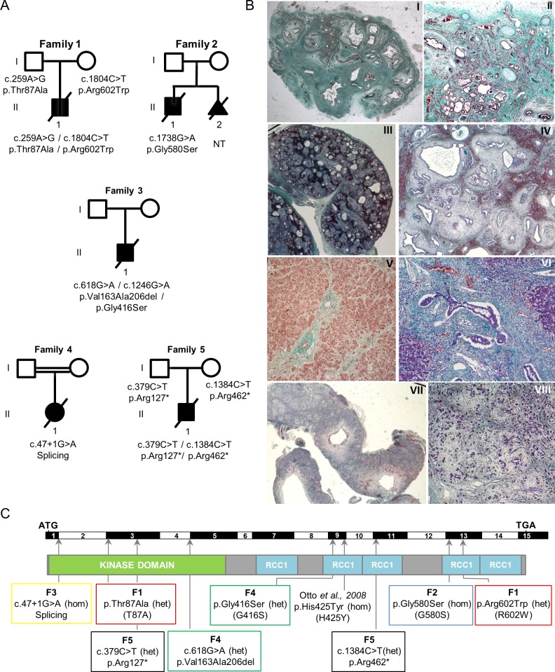
Fig 1. Identification of NEK8/NPHP9 mutations associated with severe renal cystic dysplasia.
(A) Pedigrees of the five families with identified compound heterozygous or homozygous NEK8 mutations. (B) Trichrome light green staining of kidney (I-IV), liver (V, VI) and pancreas (VII, VIII) tissue sections from individual F1 II.1 (I, II, V) and F5 II.1 (III, IV, VI-VIII). (I-II) F1 II.1 presents a major cystic hypodysplasia of the right kidney (I) and a severe dysplasia of the left kidney (II) with persistence of few differentiated nephrons between large areas of fibrosis with primitive tubules (arrowhead) and presence of metaplastic cartilage (arrows). (III-IV) Fetus F5 II.1 shows enlarged cystic kidneys (III) of apparent normal shape. High magnification (IV) revealed a diffuse architectural disorganization with the presence of cysts of various sizes, fibrosis and dysplasic tubules (asterisks) and persistence of few glomeruli (arrowhead). (V-VIII) Portal tract fibrosis with extensions bridging adjacent portal areas (arrow) and hepatocytes focally dissociated by fibrosis (V) in affected individual F1 II.1. Fetus F5 II.1 presents with massive portal fibrosis with ductular proliferation (arrows) (VI) and large cystic structures in the pancreas (VII). Higher magnification reveals a massive and diffuse fibrosis with dissociation of the pancreatic parenchyma (VIII). Magnifications are 5x (III, VII), 10x (I), 40x (II, V, VIII), 100x (IV) and 200x (VI). (C) Location of the mutations: exons, position of the start (ATG) and stop (TGA) codons, as well as the position of the kinase and five RCC1 domains are indicated. Abbreviations: heterozygous (het); homozygous (hom). The mutation reported in a patient with nephronophthisis [5] is indicated. Mutations analyzed in functional studies are indicated with one letter nomenclature.

