Figure 3.

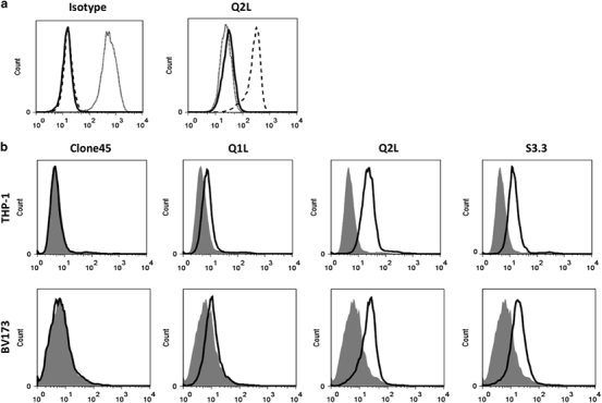
Binding of TCR-like antibodies to WT1/HLA-A0201 complexes on live cells measured by flow cytometry. (a) Binding of Q2L scFv-Fc (right) and isotype-matched TCR-like scFv-Fc (left) to T2 cells pulsed with WT1 peptide (dashed line), without peptide (solid line) or with irrelevant peptide (dotted line). T2 cells were then stained with TCR-like antibodies at 1 μg/ml, followed by fluorescent secondary antibody. (b) Recognition of the naturally presented WT1/HLA-A2 complex on tumor cells by scFv variants. The human leukemia cells, THP-1 and BV173, were stained with scFvs at 10 μg/ml, followed by fluorescent secondary antibody. Experiments from panels (a) and (b) were repeated 1–2 times with similar results.
