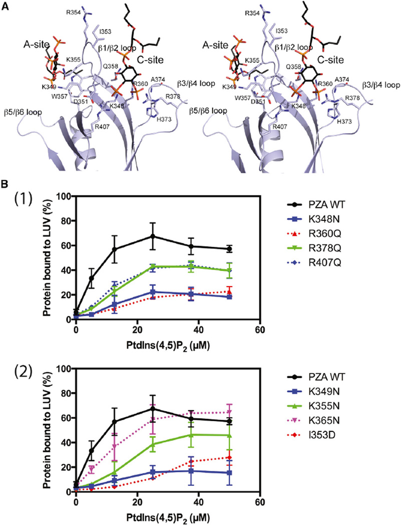
Figure 5. Mutational Analysis of Phospholipid-Binding Sites.
(A) Stereo diagram showing detailed interactions of the two bound PtdIns(4,5)P2 with residues in the binding sites. Bound PtdIns(4,5)P2 molecules are shown as stick models with carbon atoms in black, oxygen in red, and phosphor in orange. Amino acid residues taking part in interactions with PtdIns(4,5)P2 are also displayed as stick models with carbons in purple, nitrogen in dark blue, and oxygen in red. The two lipid-binding sites are also labeled as A site and C site, respectively.
(B) Binding to vesicles. (1) Mutations in canonical site. (2) Mutations in the atypical site and β1/β2 loop. Wild-type (WT) PZA and PZA with the indicated changes in amino acids were incubated with LUVs containing 15% PtdSer and the indicated concentration of PtdIns(4,5)P2.

