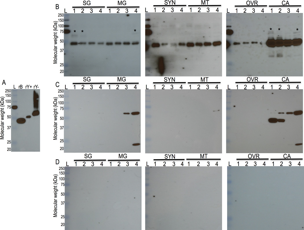
Figure 3. Western blotting analyses.
Yeast and bacteria expressed rAAS19 (Fig. 3A) and crude tick saliva protein (Figs. 3B, C, and D) extracts were subjected to western blotting analysis using the rabbit antibody to yeast expressed rAAS19 as described in materials and methods section 2.6. Figure 3A, rB = E. coli expressed rAA19, rY+ and rY− = deglycosylated and glycosylated, respectively, Pichia pastoris expressed rAAS19. Crude protein extracts of dissected salivary glands (SG), midgut (MG), synganglion (SYN), Malpighian tubules (MT), ovaries (OVR) and the remnants as carcass (CA) were subjected to western blotting analyses using antibodies to yeast expressed rAAS19 (Fig. 3B), control PBS and adjuvant injected antibody (Fig. 3C), and rabbit pre-immune serum (Fig. 3D). Lanes 1, 2, 3, and 4 = Protein extracts of unfed, 24, 72, and 120 h fed ticks respectively. L = molecular weight ladder, Asterisks marked (*) indicate potential glycosylated native AAS19 form.

