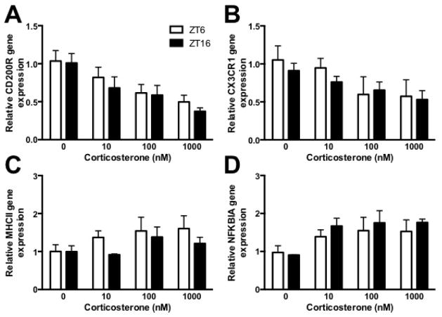
Figure 5. Microglia isolated during the middle of the light or dark phase an stimulated ex vivo with corticosterone comparably regulated gene expression.
Hippocampal microglia were isolated during the middle of the light (ZT6) or dark (ZT16) phase and treated ex vivo with 0, 10, 100, or 1000 nM corticosterone for 2h. RNA was then isolated to evaluate (A) CD200R, (B) CX3CR1, (C) MHCII and (D) NFKBIA mRNA expression. Genes are expressed relative to β actin and presented as mean ± SEM.

