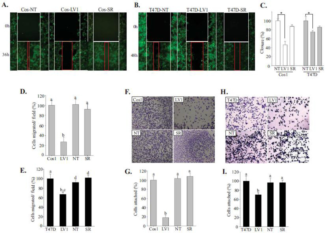
Figure 1. Shoc2 knockdown affects cell motility.
(A, B) Wound healing assay was performed using Cos1 (A) and T47D (B) cells stably expressing Shoc2 shRNA (LV1), non-targeting shRNA (NT) or expressing Shoc2 shRNA and Shoc2-tRFP (Cos-SR). The white frames indicate the width of the wound at time 0; red frames show the width of the wound 36/48h post EGF treatment.
(C) Data from independent experiments in A and B were analyzed using a Tukey Test (mean ± SD, n = 3, * p<0.001). Bars represent the change in average wound closure rates at 36/48h. The closure was calculated by measuring the average width in open area, and normalized to control cells.
(D, E) Transwell migration assay was performed in Cos1 (D) and T47D (E) cells stably expressing Shoc2 shRNA (LV1), non-targeting shRNA (NT) and Shoc2-tRFP (SR). Cells in multiple independent fields for each well were counted and three independent experiments were analyzed. Data are shown as mean ± SD; a vs. b, c vs. d, p<0.05.
(F, H) 5×10⁴ cells of Cos1 (F) and T47D (H) cells were seeded on collagen pre-coated 96-well plates for 10 min. Images of cells fixed and stained with crystal violet were obtained using a Nikon Eclipse E600 microscope.
(G, I) Cells from the experiments in F and H were solubilized with 2% SDS and subjected to colorimetric absorbance measurement (OD550). Data from three independent experiments was analyzed. Bars represent mean values ± SD, n = 3; a vs. b, p<0.05.

