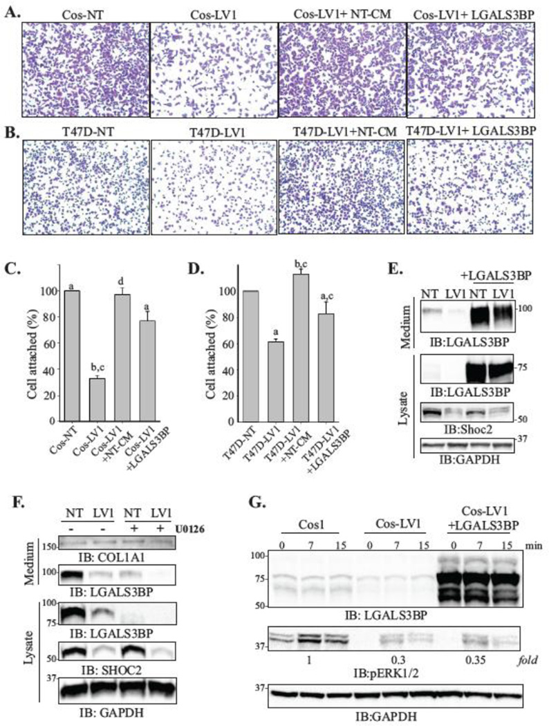
Figure 6. LGALS3BP rescues attachment of cells depleted of Shoc2.
(A, B) Cos1 (A) and T47D (B) cells (5×10⁴ cells) depleted of Shoc2 (LV1) were seeded on collagen pre-coated 96-well for 10 min. LV1+NT-CM samples were seeded in presence of cultured media from the control cells. LV1+LGALS3BP cells were transiently transfected with His-LGALS3BP 48h prior to seeding. Images of cells fixed and stained with crystal violet were obtained using Nikon Eclipse E600 microscope.
(C, D) Cells from the experiments in A and B were then solubilized with 2% SDS and subjected to the colorimetric absorbance measurement (OD550). Data from three independent experiments exemplified in A and B was analyzed (bars represent mean values ± SD; n = 3; a vs. b; c vs. d, p<0.05).
(E) Cos-NT and Cos-LV1 were transiently transfected with His-LGALS3BP. At 48h post-transfection, expression of the indicated proteins in the lysate and secretion media was analyzed using specific antibodies.
(F) Cos-NT and Cos-LV1 cells were treated with UO126 (10 uM) or DMSO for 24h at 37°C. The expression of indicated proteins in the lysate and secretion media was analyzed using specific antibodies. The data are representative of three independent experiments.
(G) Cells were transiently transfected with His-LGALS3BP. At 48h post-transfection, the cells were serum starved for 18h and stimulated with EGF (0.2 ng/ml) for 7 or 15 min. Expression of the indicated proteins was analyzed using specific antibodies.

