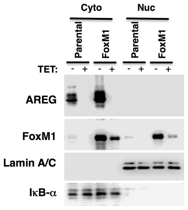
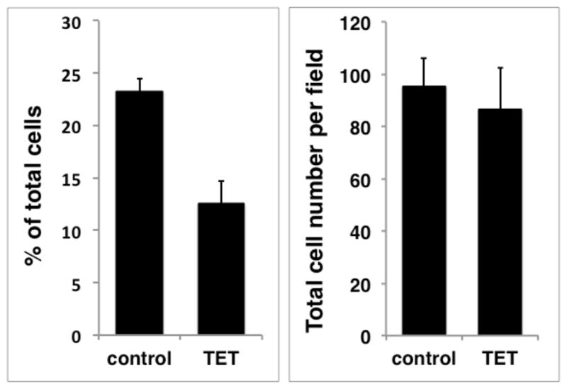
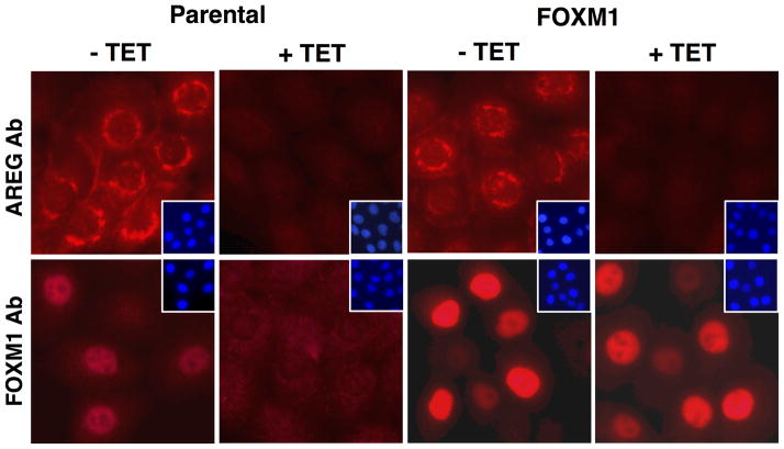
Figure 5. Constitutive expression of FoxM1 preserves nuclear and cytoplasmic FoxM1 protein in response to AREG silencing.


N/TERT-TR-shAREG keratinocytes (parental) and N/TERT-TR-shAREG keratinocytes stably infected with a constitutive FoxM1 expression construct (FoxM1-rescue cells) were cultured in KSFM medium in the presence or absence of Tet. (A) Western blots with cytoplasmic (Cyto) and nuclear (Nuc) extracts from parental and FoxM1 cells incubated for 60 h with and without Tet. Lamin A/C85 and Ikappa-B-alpha (IκB-α)86 were used as nuclear and cytoplasmic markers, respectively. (B) Immunofluorescence staining of cells incubated with and without Tet for 48 h. Insets represent DAPI staining of the same fields. Identical exposure times were used for each antibody staining and paired treatment (+/− Tet) within each cell line. (C) Quantitation of nuclear FoxM1 staining. Only cells expressing high levels of FoxM1 were scored (see Materials and Methods). Data are expressed as (left panel) percentage of strongly FoxM1-positive cells per microscopic field (10X objective, 100X final magnification); and (right panel) total cell numbers per field. Data shown represent mean and range of two experiments.

