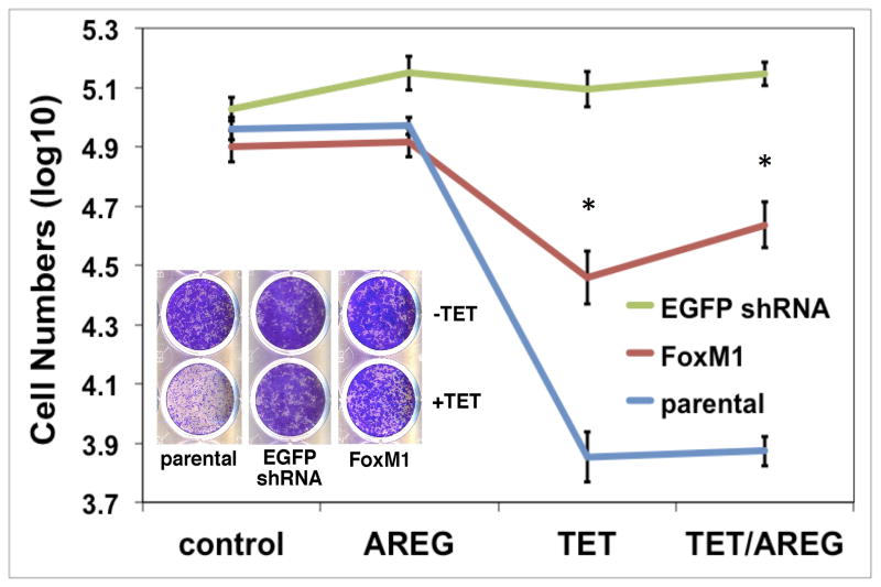
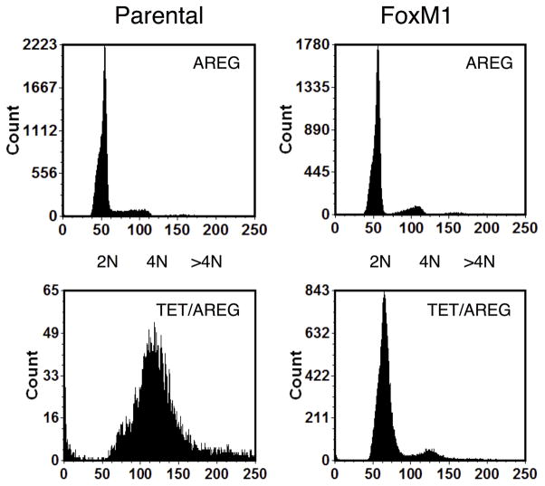
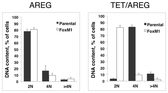
Figure 6. FoxM1 expression normalizes keratinocyte responses to cellular changes induced by AREG silencing.
N/TERT-TR-shAREG keratinocytes (parental), control keratinocytes with a Tet-inducible shRNA targeting EGFP (EGFP shRNA) and N/TERT-TR-shAREG keratinocytes stably infected with a constitutive FoxM1 expression construct (FoxM1) were subjected to six day growth assays in the presence or absence of 1 μg/ml Tet with and without 100 ng/ml recombinant human AREG. (A) Cell numbers were assessed by flow cytometry based cell counting with fluorescent reference beads. Data are expressed as log(10) of total cell numbers for the four different treatments that are indicated underneath the x-axes. Data represent keratinocyte cell numbers per cm2 of growth area, and represent mean +/− within-subject SE computed by the Cousineau-Morley method, n=4 for the parental and FoxM1 cell lines, n=3 for EGFP shRNA cell line, with three biological replicates per “n”. The inset shows a crystal violet staining of a typical result. Significant pairwise contrasts (mean ratio; corrected P): Tet treatment: FoxM1 vs. parental (4.0; 0.029), EGFP shRNA vs. parental (17.4; 0.00096), EGFP shRNA vs. FoxM1 (4.3; 0.027); Tet/AREG treatment: FoxM1 vs. parental (5.8; 0.0040), EGFP shRNA vs. parental (18.8; 0.00012), EGFP shRNA vs. FoxM1 (3.2; 0.032). (B) Cell cycle distribution of DAPI-stained keratinocytes at the end of six-day growth assays. (C) Quantitation of cell cycle distributions for all experiments. Data are expressed as % of cells with 2N, 4N or > 4N DNA content, mean +/− standard deviation, n=4.


