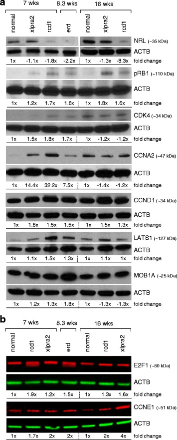
Fig. 6.

Western blot analysis of normal, xlpra2, rcd1, and erd retinas. a Blots exposed on autoradiograph films; b Li-COR immunoblotting system. The band intensity normalized to the actin density of the normal control (7 wks for ages 7 and 8.3 wks; 16 wks for 16 wk mutant samples) was set to one, and fold changes were calculated and shown below each of the blots
