
Fig. 2.
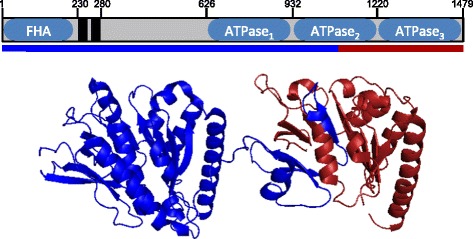
Domain architecture of EssC protein. Sequence variable region of EssC shown in red on the X-ray structure of the C-terminal ATPase domains of G. thermodentrificans EssC. The blue and red line underneath the linear representation of EssC marks the extent of the conserved and variable regions identified in the comparison between the essC1 and essC2 illustrated in Fig. 1