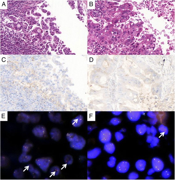
Fig. 3.

ALK rearrangement status and ALK expression determined by fluorescence in-situ hybridization (FISH) and immunohistochemistry (IHC) in two regions of the same primary lung tumor. a, b examination after hematoxylin-eosin-safran (HES) staining (×400): histological features of adenocarcinoma from the bronchoscopy (a) and the surgical resection (b) specimens. c, d ALK expression: very faint positive immunoreactivity (score, 0/1) in both the small biopsy and the corresponding surgical resection specimen. e, f Typical break-apart pattern observed by FISH (arrow): 25 % of rearranged tumor nuclei detected in the biopsy sample, versus 2 % in the excision specimen
