
Figure 1.
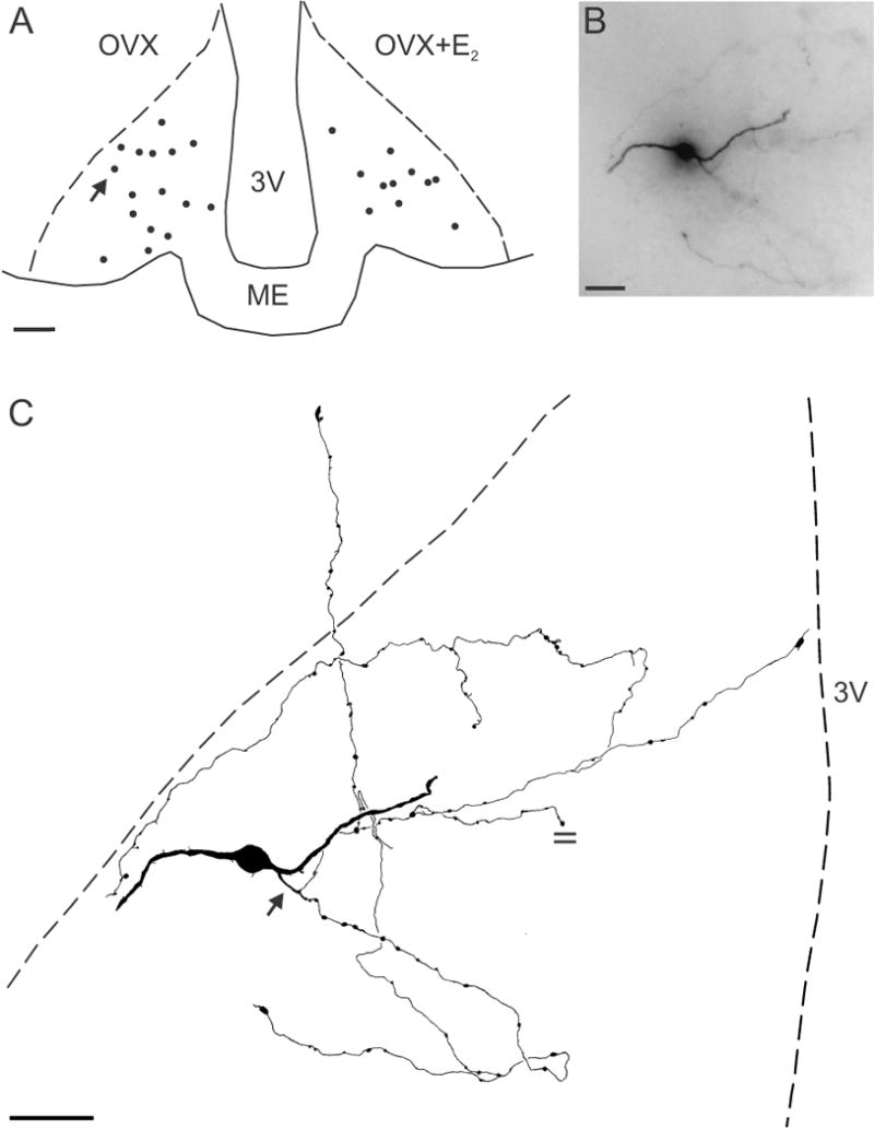
(A) Computer-assisted map showing the location of biocytin-filled cell bodies at the mid-level of the arcuate nucleus in OVX (left) and OVX+E2 (right) Tac2-EGFP mice. The arrow in A shows the location of the neurone illustrated in B and C. (B) Photomicrograph of a biocytin-filled neurone from an OVX animal reveals a soma (out of focus), two dendrites and an axon arbor out of the focal plane. (C) Camera lucida drawing of the neurone shown in B. This KNDy neurone was spiny with two primary dendrites. The axon (arrow) emerges from a proximal dendrite and gives rise to extensive local collaterals with en passant and terminal boutons within the arcuate nucleus. One axonal branch terminates within the ependymal layer. The parallel lines mark an axon cut at the surface of the section. Scale bar in A = 100 μm, B = 20 μm and C = 50 μm, ME, median eminence, 3V, 3rd ventricle.