
Figure 3.
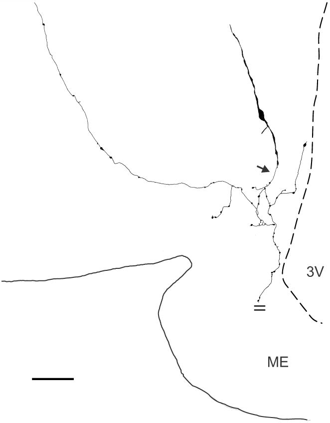
Camera-lucida drawing of a biocytin-filled KNDy neurone from an OVX + E2 Tac2-EGFP mouse (mid-level arcuate). This neurone is sparsely-spined with two primary dendrites. A beaded axon emerges ventrally (arrow) from a distal dendrite and sends collaterals that terminate within the arcuate nucleus, the ependymal layer of the 3rd ventricle and a dorsolateral branch extending beyond the borders of the arcuate nucleus. An axon collateral extending to the median eminence was cut (parallel lines) at the surface of the tissue slice. Scale bar = 50 μm. ME, median eminence, 3V, 3rd ventricle.