
Figure 5.
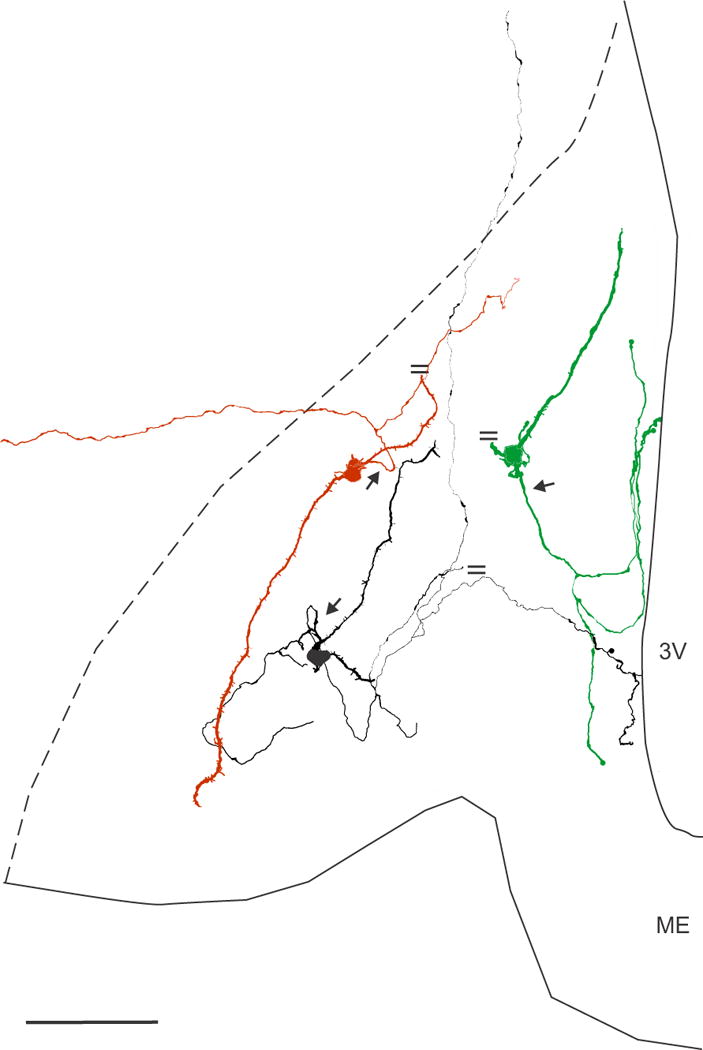
Neurolucida reconstruction of 3 biocytin-filled KNDy neurones in the arcuate nucleus of OVX Tac2-EGFP mice. These neurones digitized from three separate mice, had simple dendritic fields that were restricted to the arcuate nucleus, but different axonal trajectories. The parallel lines indicate cut processes. The axon (arrow) of the red neurone emerged from a proximal dendrite, bifurcated and sent one branch laterally outside of the arcuate nucleus. One of the dendrites was cut at the surface of the section. The axon of the black neurone emerged from a proximal dendrite (arrow) and arborized locally within the arcuate nucleus. One branch extended dorsally to terminate in an axonal arbor within the dorsomedial nucleus (not shown). One axon was cut at the surface of the section and another branch extended to the wall of the third ventricle. The green neurone had a dendrite that was truncated at the surface of the tissue section and an axon (arrow) emerging from the cell body with branches terminating in the arcuate nucleus and projecting to the ependymal lining. Scale bar = 100 μm. 3V, 3rd ventricle, ME, median eminence.