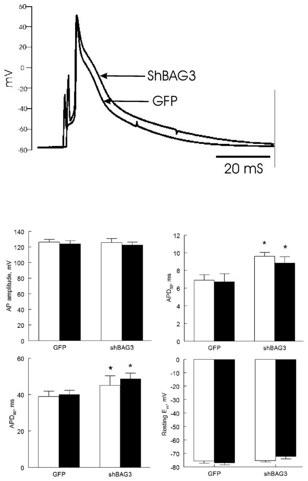
Figure 5. BAG3 downregulation prolongs action potential duration (APD).
(A). Representative action potentials of GFP and shBAG3 myocytes are shown. (B). Means ± SE of resting membrane potential (Em), action potential amplitude, and APD at 50% (APD50) and 90% repolarization (APD90) from 7 GFP (3 mice) and 5 shBAG3 myocytes (3 mice), both before (open bars) and after (filled bars) 1 μM Iso are shown. *p<0.045; GFP vs. shBAG3.

