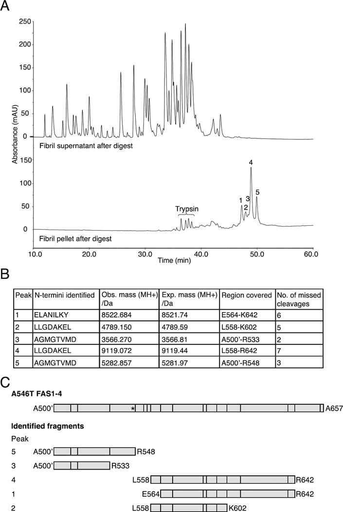
Figure 4.
Isolation of trypsin-generated fragments that represent the fibril core region. Fibrillated A546T FAS1-4 was treated with trypsin at a ratio of 1:1 w/w and separated into pellet and supernatant fractions. A, Chromatograms from RP-HPLC of the pellet (lower chromatogram) and supernatant (upper chromatogram) fractions. Five peaks (marked 1–5), representing the fibril core, were specifically retained in the pellet fraction, and four minor peaks corresponded to peptides from the high amount of trypsin added (data not shown). B, N-terminal sequencing and mass spectrometry analyses determined the sequences of the five peptides that covered the fibril core region. The table contains the identified N-termini, observed masses, expected masses, regions covered in A546T FAS1-4, and the number of missed trypsin cleavages. C, Schematic representation of the identified fibril core fragments with vertical lines representing potential trypsin cleavage sites and an asterisk marking the A546T mutation site.

